
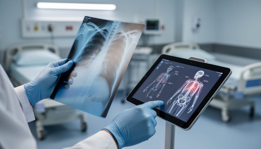
Musculoskeletal imaging refers to the medical imaging techniques used to visualize bones, joints, muscles, ligaments, tendons, and other connective tissues in your body. These diagnostic tools help doctors identify fractures, arthritis, sports injuries, degenerative conditions, and other problems affecting your skeletal and muscular systems. For men in particular, imaging plays a crucial role in catching early signs of osteoporosis, diagnosing chronic joint pain, evaluating sports-related injuries, and monitoring conditions that become more common after age 40.
Understanding the fundamentals of musculoskeletal imaging matters because you’ll likely encounter at least one of these procedures during your lifetime, whether after a weekend hockey injury, to investigate persistent back pain, or as part of routine screening for bone health. Knowing which imaging method your doctor might order, what each one reveals, and how to prepare can reduce anxiety and help you participate more actively in your care decisions.
This guide breaks down the most common imaging modalities, from standard X-rays to advanced MRI and CT scans, explaining in straightforward terms how each works, what conditions they’re best suited to detect, and when your healthcare provider might recommend one over another. You’ll also learn how imaging fits alongside other diagnostic tools and lifestyle factors, including bone-supporting nutrients to maintain long-term musculoskeletal health. The goal is simple: give you the knowledge to understand what’s happening when imaging is recommended and what those results mean for your wellness journey.
What Musculoskeletal Imaging Means
Musculoskeletal imaging refers to a set of medical visualization techniques that allow healthcare providers to see inside your body and examine bones, joints, muscles, tendons, and ligaments without surgery. Think of it as taking photographs of your skeleton and surrounding tissues using specialized technology, from traditional X-rays to advanced MRI machines, to identify problems that aren’t visible from the outside.
- Musculoskeletal System
- The framework of bones, joints, muscles, tendons, and ligaments that supports your body, enables movement, and protects vital organs.
- Diagnostic Imaging
- Medical tests that create visual representations of internal body structures to help doctors identify diseases, injuries, and abnormalities.
- Bone Density
- The measurement of mineral content in bones, indicating their strength and resistance to fractures.
- Soft Tissue
- Non-bony structures including muscles, tendons, ligaments, cartilage, and fat that support and connect the skeletal system.
- Radiology
- The medical specialty focused on using imaging technologies to diagnose and sometimes treat diseases and injuries.
What sets imaging apart from other diagnostic methods is its ability to provide direct visual evidence of structural problems. While a physical examination might reveal tenderness or limited range of motion, imaging shows the actual fracture line, cartilage wear, or tendon tear causing those symptoms. Blood tests can indicate inflammation, but an X-ray or MRI pinpoints exactly where arthritis has damaged a joint.
For Canadian men, this distinction matters significantly. Musculoskeletal imaging catches fractures from weekend hockey games, reveals early-stage osteoporosis before bones become dangerously fragile, identifies rotator cuff tears that explain persistent shoulder pain, and tracks arthritis progression in weight-bearing joints. It transforms vague discomfort into concrete diagnoses, replacing guesswork with clear visual documentation that guides treatment decisions and prevents minor issues from becoming major disabilities.
How Musculoskeletal Imaging Works
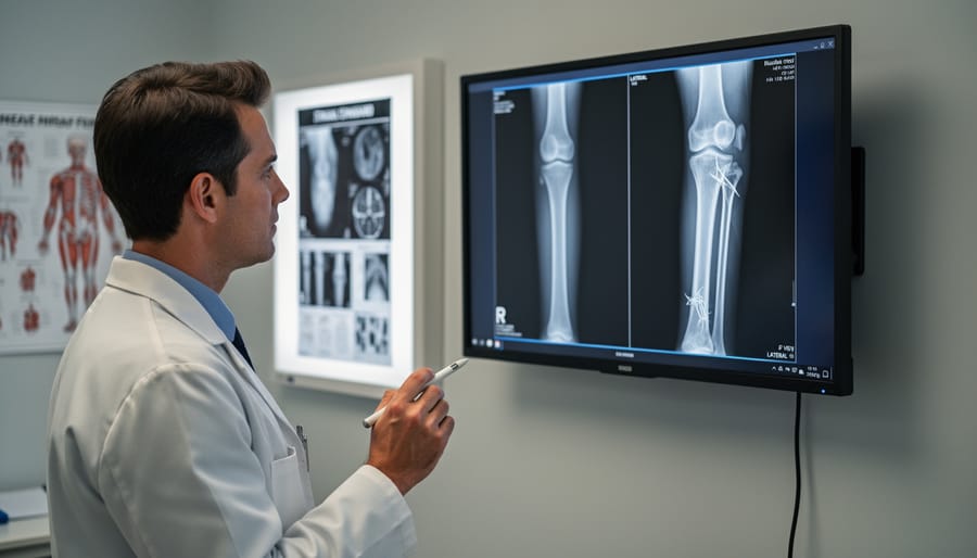
Musculoskeletal imaging works by directing different forms of energy through your body to create detailed pictures of structures that doctors can’t see or feel during a physical exam. Each imaging technique uses a specific type of energy, whether X-ray beams, magnetic fields, or sound waves, that interacts with your tissues in predictable ways. These interactions produce signals that specialized equipment captures and converts into visual images, revealing the shape, density, and condition of your bones, joints, and soft tissues.
During a typical imaging procedure, you’ll be positioned so the area of concern sits between the energy source and the detector. For X-rays, this means standing or lying while a brief burst of radiation passes through your body, dense structures like bones block more radiation and appear white on the image, while softer tissues let radiation through and show up as darker shades. The entire process usually takes just minutes and causes no sensation.
More sophisticated technologies like MRI work differently. Instead of radiation, an MRI machine uses powerful magnets to align hydrogen atoms in your body, then measures how these atoms respond when radio waves disturb that alignment. Different tissues respond at different rates, creating contrast that reveals not just bones but also cartilage, ligaments, tendons, and even subtle inflammation. The trade-off is time, MRI scans often require 30 to 60 minutes of holding still inside the machine.
Once captured, these images land on a radiologist’s screen. These specialized physicians analyze the images systematically, comparing what they see to normal anatomy and looking for signs of injury, disease, or degeneration. They document their findings in a detailed report that your primary care provider or specialist uses to confirm diagnoses, plan treatment, or decide whether additional imaging is needed. The technology itself is passive, it simply provides the visual information that expertise then interprets.
Main Types of Musculoskeletal Imaging
X-Rays (Radiography)
X-rays remain the workhorse of musculoskeletal imaging because they deliver clear bone images quickly and affordably. When you experience a suspected fracture, joint pain, or mobility issues, an X-ray is typically the first diagnostic step your doctor orders. The procedure takes only minutes, you position the affected area against a detector plate while a controlled burst of radiation passes through your body, creating a detailed shadow image where dense bone appears white and softer tissues show as darker shades.
This imaging method excels at revealing bone fractures, stress fractures, joint misalignment, and signs of arthritis like bone spurs or narrowed joint spaces. For men recovering from sports injuries or monitoring conditions affecting strength and recovery X-rays provide baseline assessments and track healing progress over time. Most Canadian clinics and emergency departments have X-ray capabilities, meaning you’ll rarely wait long for this essential diagnostic tool. The radiation exposure is minimal, roughly equivalent to a few days of natural background radiation, making X-rays a safe, practical choice for initial bone and joint evaluation.
CT Scans (Computed Tomography)
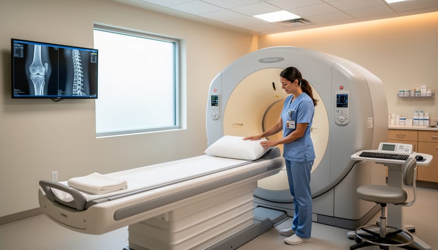
CT scans use X-ray technology combined with computer processing to create detailed cross-sectional images of your bones and joints, think of it as looking at your skeleton in thin slices rather than as a single flat picture. This layered view reveals fracture patterns, bone alignment issues, and structural details that standard X-rays can’t capture.
For men dealing with complex injuries, CT excels at showing exactly how bone fragments have shifted after a fracture, which helps orthopedic surgeons plan precise repairs. It’s particularly valuable for assessing injuries to areas with complicated anatomy like the pelvis, spine, or wrist where bones overlap on regular X-rays.
CT also detects subtle bone tumors and evaluates bone density distribution, making it useful when doctors suspect conditions beyond simple breaks. The procedure itself is quick, typically five to ten minutes, and requires you to lie still while the scanner rotates around the targeted area.
The trade-off? CT delivers higher radiation exposure than plain X-rays, so physicians order it when the diagnostic benefits clearly outweigh this consideration. You won’t need CT for routine bone health checks, but when complexity demands clarity, it provides the detailed roadmap your healthcare team needs.
MRI (Magnetic Resonance Imaging)
MRI stands out as the most powerful tool for examining soft tissues that X-rays simply can’t capture. Unlike radiography or CT, MRI uses strong magnetic fields and radio waves to create detailed images of muscles, tendons, ligaments, cartilage, and spinal discs without any radiation exposure.
For Canadian men dealing with persistent joint pain or sports injuries, MRI often provides the definitive diagnosis. It excels at revealing rotator cuff tears in the shoulder, meniscus damage in the knee, and herniated discs in the spine. The technology can show inflammation, partial tears, and subtle structural changes that explain ongoing symptoms even when other imaging appears normal.
An MRI scan typically takes 30 to 60 minutes. You’ll lie still inside a tunnel-shaped machine that makes loud knocking sounds (earplugs help). The machine’s magnetic field is completely safe, but you must remove all metal objects beforehand. Some facilities offer open MRI units for those who feel claustrophobic.
The main drawback is accessibility. MRI machines are expensive to operate, which means longer wait times through the public system and higher costs if you pay privately. However, when soft tissue detail matters for diagnosis or surgical planning, MRI remains unmatched.
Ultrasound
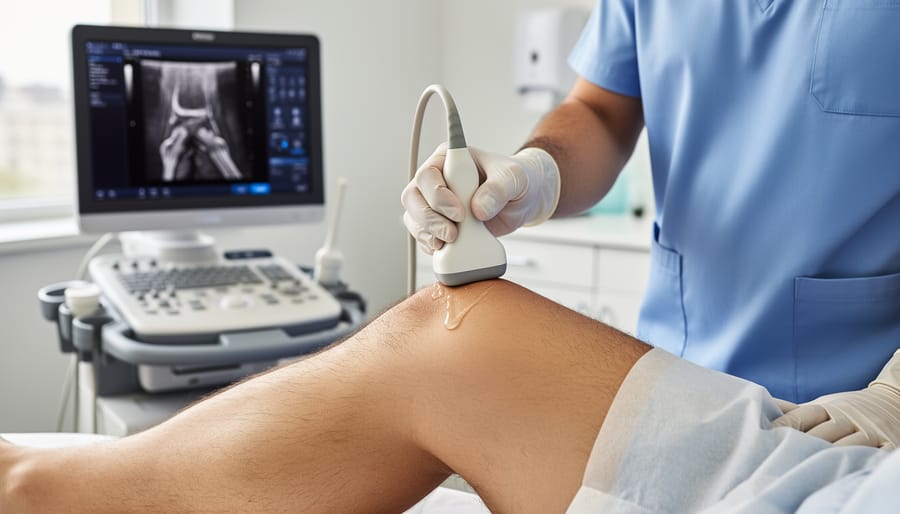
Ultrasound imaging uses high-frequency sound waves to create real-time images of soft tissues beneath the skin. A handheld device called a transducer sends sound waves into your body, which bounce back at different rates depending on the tissue they encounter. The machine translates these echoes into moving images displayed on a screen, allowing the technician or doctor to view structures as they shift and move.
For musculoskeletal concerns, ultrasound excels at examining tendons, muscles, ligaments, and bursae, tissues that often don’t show clearly on X-rays. It’s particularly valuable for diagnosing rotator cuff tears, tennis elbow, Achilles tendon injuries, and muscle strains. Because the images update in real time, healthcare providers can watch how structures move during certain motions, revealing dynamic problems that static imaging might miss.
Canadian men appreciate ultrasound’s accessibility. The equipment is portable, so it’s available in many clinics without requiring a hospital visit. There’s no radiation exposure, making it safe for repeated use when monitoring healing progress. Doctors also use ultrasound guidance to precisely place injections into joints or around tendons, improving treatment accuracy. The procedure takes 15 to 30 minutes and involves only gel and gentle probe pressure against your skin.
DEXA Scans (Bone Density Scans)
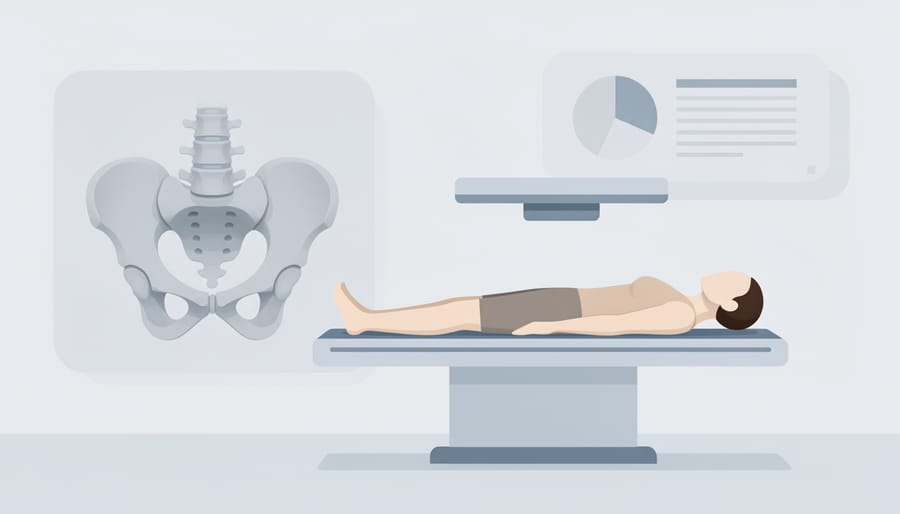
DEXA scans, short for dual-energy X-ray absorptiometry, use two different X-ray beams to measure how much mineral is packed into your bones. The machine compares the energy absorption between the two beams as they pass through bone tissue, calculating your bone mineral density (BMD) with remarkable precision. This technique has become the gold standard for diagnosing osteoporosis because it can detect even small changes in bone density that other imaging methods miss.
For Canadian men, DEXA scans are particularly valuable after age 65 or earlier if you have risk factors like low testosterone, long-term corticosteroid use, or a family history of fractures. The test takes about 10 to 20 minutes and involves lying still on a padded table while a scanner arm passes over your lower spine and hip, the areas most prone to osteoporotic fractures. Unlike other imaging, DEXA uses extremely low radiation doses, about one-tenth of a standard chest X-ray.
Your results come as a T-score comparing your bone density to that of a healthy 30-year-old. A score above -1 is normal, between -1 and -2.5 indicates osteopenia (low bone mass), and -2.5 or below confirms osteoporosis. These numbers help your doctor assess your fracture risk and determine whether you need medication or lifestyle changes to protect your skeletal health.
Nuclear Medicine Bone Scans
Nuclear medicine bone scans inject a small amount of radioactive tracer into the bloodstream, which accumulates in areas where bone cells are actively rebuilding or repairing. Special cameras then detect these “hot spots” of metabolic activity. This makes bone scans find hidden injuries like stress fractures, infections, or tumors that don’t appear on X-rays or CT scans, especially in their early stages. The procedure typically takes two to four hours, including waiting time for the tracer to circulate, and is particularly valuable when doctors suspect problems affecting multiple bone sites simultaneously.
When Musculoskeletal Imaging Is Used
Diagnosing Acute Injuries
When you twist an ankle during a pickup basketball game, fracture your wrist in a fall, or tear a muscle during weightlifting, musculoskeletal imaging provides the definitive answer about what happened inside. X-rays are typically the first choice for suspected bone injuries, they quickly reveal fractures, show whether bones are properly aligned, and help determine if a dislocation occurred. A radiologist can usually spot a break within minutes, allowing your healthcare provider to decide on immediate treatment.
For soft tissue injuries like ligament tears or muscle strains, MRI becomes the go-to option. If your knee buckles and swells after a sports collision, an MRI can visualize whether you’ve torn your ACL, damaged your meniscus, or simply strained surrounding tissues. CT scans fill the gap when fractures are complex or involve joints, they provide three-dimensional detail that helps orthopedic surgeons plan repairs.
Ultrasound serves acute scenarios too, particularly for examining shoulder rotator cuff tears or calf muscle injuries where real-time imaging helps assess the extent of damage. The choice of imaging depends on your symptoms, the injury mechanism, and what structures your doctor suspects are affected.
Monitoring Chronic Conditions
Musculoskeletal imaging doesn’t end with diagnosis, it’s equally vital for tracking chronic conditions over months and years. For men living with osteoporosis, repeat DEXA scans typically occur every one to two years to measure whether bone density is improving, stabilizing, or declining. This data helps physicians adjust medication, supplement dosages, and lifestyle recommendations before fractures occur.
Arthritis monitoring relies on periodic X-rays or MRI scans to document joint space narrowing, cartilage loss, and inflammation patterns. Seeing objective evidence of progression, or stability, guides decisions about changing medications, adding physical therapy, or considering surgical options like joint replacement. Men with rheumatoid arthritis may undergo imaging every six to twelve months to catch early joint damage that might not yet cause obvious symptoms.
Treatment effectiveness becomes measurable through imaging. After starting osteoporosis medication, subsequent scans reveal whether the drug is preventing further bone loss. Post-surgery imaging confirms proper healing, hardware placement, and absence of complications. This visual feedback transforms chronic condition management from guesswork into evidence-based care, giving both patient and provider concrete markers of progress.
Preventive Screening
Most men don’t think about musculoskeletal imaging until something hurts. Yet preventive screening can catch problems before they cause fractures or disability, particularly important as bone loss happens silently.
Canadian men should consider bone density screening starting around age 65, or earlier if risk factors exist. These include family history of osteoporosis, previous fractures from minor falls, long-term corticosteroid use, low testosterone, smoking, excessive alcohol consumption, or conditions like rheumatoid arthritis and inflammatory bowel disease. Men with significant weight loss or chronic diseases affecting calcium absorption benefit from earlier baseline scans.
A DEXA scan takes about 15 minutes and involves no needles or special preparation. It measures bone mineral density at the hip and spine, the sites most vulnerable to fractures. Results determine whether your bones are normal, show early loss (osteopenia), or indicate osteoporosis requiring treatment.
Unlike mammograms or colonoscopies, bone density screening isn’t routinely promoted for men, despite roughly one in four Canadian men over 50 eventually experiencing osteoporotic fractures. Proactive screening identifies thinning bones when lifestyle changes, calcium, vitamin D, and weight-bearing exercise can still make significant protective differences.
What to Expect During Musculoskeletal Imaging
Walking into an imaging appointment for the first time can feel uncertain. Knowing what happens during common musculoskeletal imaging procedures helps reduce anxiety and ensures you’re prepared.
X-rays are the quickest and simplest. You’ll be positioned near the X-ray machine, standing, sitting, or lying down depending on the body part being examined. A technologist will place your limb or joint in specific positions, sometimes asking you to hold still for a few seconds while images are captured. The entire process typically takes 5-10 minutes. You can wear most clothing, though you may need to remove metal objects like watches or jewelry.
CT scans involve lying on a motorized table that slides through a large, doughnut-shaped scanner. The machine rotates around you, taking multiple images from different angles. You’ll hear whirring or clicking sounds, but the scan itself is painless. Most musculoskeletal CT scans take 10-15 minutes. Sometimes contrast dye is injected through an IV to improve image clarity.
MRI scans require you to lie still inside a narrow tube for 30-60 minutes. The machine produces loud thumping and buzzing noises, you’ll be offered earplugs or headphones. Because MRIs use powerful magnets, you must remove all metal items beforehand. Some people feel claustrophobic; if this concerns you, mention it when booking, sedation or open MRI options may be available.
Ultrasound examinations are the most interactive. You’ll lie on an examination table while a technologist applies gel to your skin and moves a handheld probe over the area being examined. The process is painless and typically takes 15-30 minutes.
DEXA scans are straightforward: you lie flat on a padded table while a scanning arm passes over your body, usually focusing on your spine and hips. The scan is completely painless and takes about 10 minutes.
For all procedures, arrive a few minutes early to complete paperwork. Most imaging centers are cool, so bring a sweater. Results typically reach your doctor within a few days.
Common Questions About Musculoskeletal Imaging
Most Canadian men want straightforward answers before stepping into an imaging clinic. Here are the questions that come up most often, along with practical guidance to help you feel prepared.
Is radiation from X-rays and CT scans dangerous?
The radiation dose from a single X-ray or CT scan is very small and considered safe for diagnostic purposes. Modern equipment uses the lowest effective dose possible, and the diagnostic benefits far outweigh the minimal risk for most men.
How long until I get my imaging results?
Turnaround times vary by urgency and facility. Emergency fractures may be read within hours, while routine screening like bone density scans typically take three to seven business days for a radiologist’s report to reach your doctor.
Are musculoskeletal imaging procedures covered by provincial health insurance?
Most medically necessary imaging ordered by your physician is covered under provincial health plans across Canada, though some provinces may have different coverage for specific scans. DEXA scans for osteoporosis screening may require meeting age or risk criteria.
What if I’m claustrophobic and need an MRI?
Tell your healthcare provider beforehand, many facilities offer open MRI machines with more space, or you may be prescribed mild anti-anxiety medication for the procedure. Music or headphones during the scan can also help.
How often should men get bone density scans?
Canadian guidelines typically recommend men start screening at age 65, or earlier if you have risk factors like family history, previous fractures, or long-term steroid use. Follow-up scans are usually done every two years to monitor changes.
Beyond these core musculoskeletal questions men also wonder whether they can request a specific imaging type. While you can certainly discuss preferences with your doctor, the imaging method chosen depends on what needs to be seen, your physician will select the most appropriate technique based on your symptoms and the diagnostic goal.
Wait times for non-urgent imaging vary significantly across provinces and between urban and rural areas. If you’re concerned about delays, ask your doctor’s office about typical timelines at local facilities or whether there are alternative clinics with shorter waits. Private imaging clinics exist in some provinces, but you’ll pay out-of-pocket unless medically necessary public options have unreasonable delays.
Musculoskeletal imaging isn’t something to dread, it’s one of the most valuable tools you have for protecting your bone and joint health as you age. Whether you’re dealing with a sudden injury, managing a chronic condition like arthritis, or simply want to understand your fracture risk, these imaging techniques provide the clear answers your healthcare team needs to guide effective treatment and prevention strategies.
For Canadian men, integrating imaging into your health routine can make the difference between catching osteoporosis early and facing preventable fractures later. It transforms skeletal wellness from guesswork into measurable data, helping you and your men’s wellness doctor make informed decisions about exercise, nutrition, and medication.
The key is knowing when imaging makes sense for your situation. Not every ache requires an MRI, but ignoring persistent symptoms or skipping recommended bone density screening can leave serious issues undetected. Talk with your healthcare provider about your family history, activity level, and age-related risk factors to determine which imaging, if any, belongs in your preventive care plan.
Ultimately, musculoskeletal imaging demystifies what’s happening beneath the surface. It empowers you to take charge of your skeletal health with confidence rather than anxiety, turning uncertainty into actionable knowledge that keeps you moving well for years to come.